


REMY: HEAD-FIXED BEHAVIOR AND ELECTROPHYSIOLOGY IN RATS
In collaboration with the Totah lab at the University of Helsinki, we are developing REMY, a set of tools that enable complex behavioral experiments with head-fixed, behaving rats on a treadmill/VR setup. We are also modifying those tools to enable awake brain imaging in MRI scanners.
Head fixation methods for mice freely running on a wheel/treadmill have proven revolutionary in systems neuroscience (e.g. Dombeck et al 2010, Saleem et al. 2013 Poort et al. 2022). They provide the necessary stability to utilize cutting-edge technologies for recording neuronal activity while animals perform behavioral tasks, including in virtual reality environments. Head fixation also enables more accurate ways to monitor eye movements and facial expressions (for experimental and welfare purposes), and better control over the animal's sensory input, when compared to freely moving experiments.
Rats are capable of more complex behaviors while enabling higher–density neuronal recordings than mice. Yet due to their size and the strength that comes with it, head fixation has so far required highly restrictive fixation methods that prevent the animal from moving, let alone running naturally in a virtual reality environment.
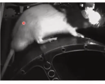
High-level concept of the rat head-fixed behavior setup
REMY changes that. The core innovation is a headplate - in biocompatible material - that secures a strong and stable head-fixation, removing the need for skull screws in the process. It was first validated for chronic electrophysiology recordings (EEG) during a Go/No-Go auditory-visual discrimination task (preprint) and during a study of the brightness illusion response (paper). Both studies had animals running freely on a wheel. Current studies in the Totah lab now employ flexible high-density probes with similar behavioral tasks.
We are now working on expanding the range of capabilities to acute electrophysiology recordings and calcium imaging, as well as MRI compatibility for the head fixation element, to enable fMRI brain imaging in awake animals.
Performance
Rat head fixation was successfully achieved by combining an innovative headplate design with a carefully adapted surgery protocol.
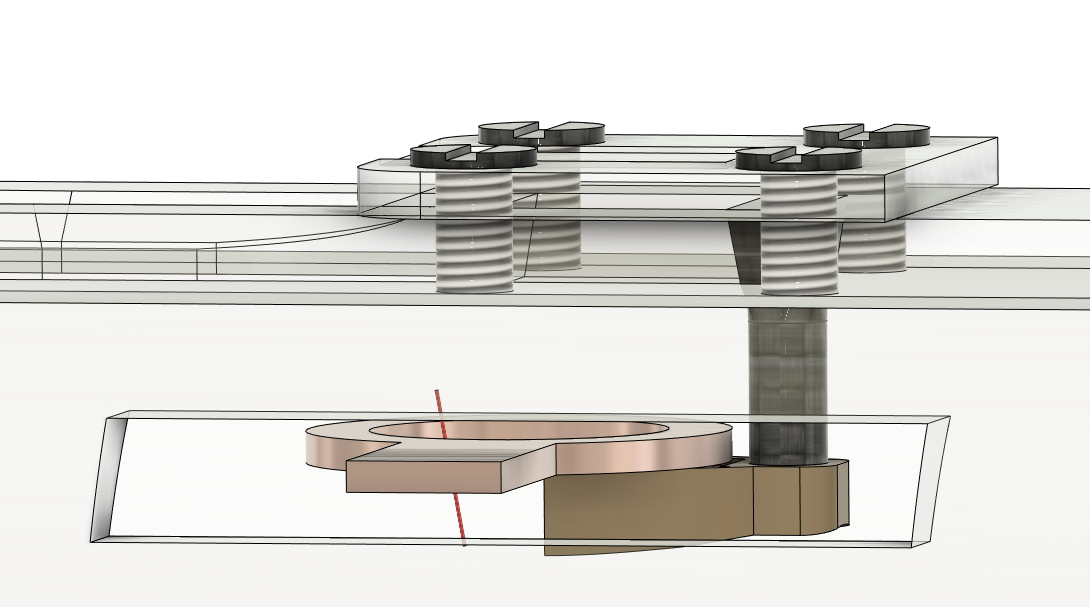
Unprecedented skull stability - These advances in design and methods allow an implant so stable that skull screws are unnecessary.
Train head-fixed behavior first, implant later - The headplate leaves most of the skull free, and after implantation can be filled with silicon (that's why we call it a chamber) and sealed with a plastic cap. The animal can then be head-fixed and trained, and once the task is performed to a chosen criterion, a craniotomy can be performed and your recording technology of choice implanted.
Developed over a period of several years, the method was thoroughly tested:
125 animals implanted
93% success rate
3-8 months of recordings from behaving animals
Validated for chronic ECoG arrays and silicon probes
Related publications
Data collected on behaving head-fixed rats has so far led to several publications, some of which have already been through peer-review:
- Vasilev et al. Brightness illusions evoke pupil constriction and a preceding primary visual cortex response in rats. Cerebral Cortex 13:12 (2023) https://doi.org/10.1093/cercor/bhad090
Multiple preprints and 2 posters presented at SfN 2022:
- Klein et al. Aberrant prefrontal activity and arousal level correlate with action initiation and response vigor.
- Figueira et al. Volitional stopping is preceded by a transient beta oscillation.
- Vasilev et al. Focusing perceptual attention in one modality constrains subsequent learning in another modality. bioRxiv 2022.01.22.477334; doi: https://doi.org/10.1101/2022.01.22.477334
- Poster 235.05 A transient beta oscillation occurs with high temporal regularity prior to stopping an ongoing movement
- Poster 565.01 Distinct anterior cingulate neurons drive changes-of-mind and monitor past performance
A methods paper describing all procedures is in preparation.
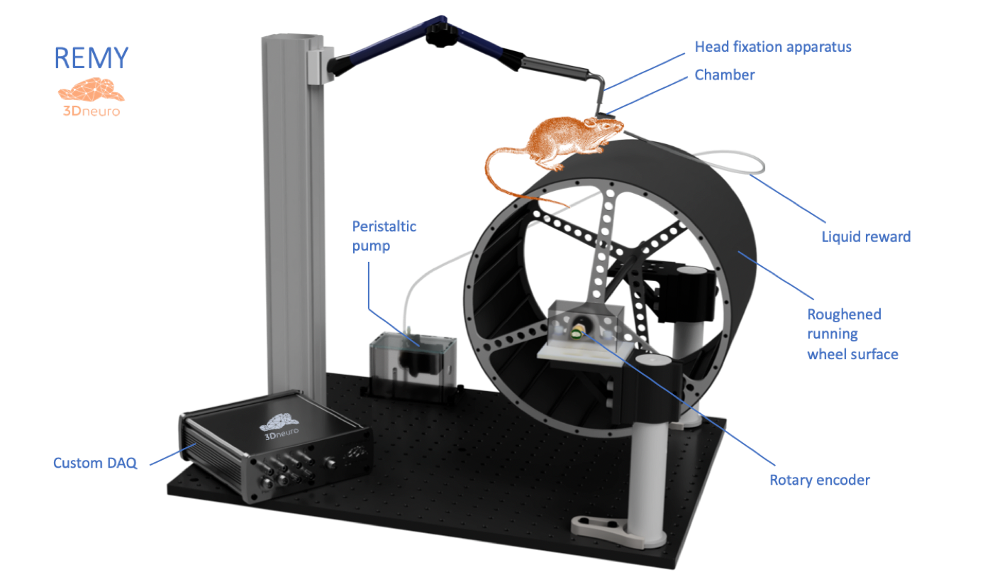
Elements of the rat head-fixed behavior setup
In addition to the head fixation itself, the setup comprises several other components:
- A cylindrical treadmill with a roughened fiberglass surface. This provides appropriate traction for the animal’s paws. Running speed is measured by a rotary encoder.
- A liquid reward delivery system.
- A metal frame, based on an optical rail system, that can flexibly accommodate various sensors (cameras to monitor the animal and measure eye movements), actuators (sensory stimulation with sound, etc) and accessories (e.g. infrared lighting).
- A microcontroller that interfaces the various sensors, actuators and accessories using analog and digital input/output.
- Grounding.
REMY: MRI-compatible head fixation
Current status