


Background
In addition to microdrives, chronic implants with silicon probes require a protective structure (head gear). The head gear serves multiple purposes:
- Protect the implant site and microdrives/probes from animals and debris, by providing a physical barrier
- Hold the high-density connectors (or headstages, for Neuropixels and Cambridge Neurotech mini amps) and decouple them physically from the microdrives (in the following, we will use Omnetics connector, which are still the most common ones, as a blanket term for all high density connectors, head stages, or EIBs that are the interface between the flex cable on the probe and the tether cable leading to your recording system)
- (optionally) Improve recorded signal by acting as a Faraday cage (less important for the nowadays common neural data acquisition systems that digitize the signal very close to the electrode)
- (optionally) Provide possibility for head fixation of the animals
The head gear that is typically used with the R2Drive and 3Drive is based on a protective copper mesh construction originally by Vandecasteele et al [1]. This solution (which we just refer to as copper mesh) is still the gold standard, as it allows for a very flexible way of realizing even complex implants with multiple drives (see, for example, [2, 3] who implanted three 3Drives simultaneously using this approach). No matter which of our head gear solutions you choose, we do recommend you familiarize yourself with the original JoVE protocol, as it helps in understanding the purpose and underlying ideas of the system.
However, the flexibility of the copper mesh comes at the cost of complexity – the implant has to be built/finalized during surgery, extending surgery times by up to an hour (for experienced users) or more. In addition, the Omnetics connectors are secured onto the copper mesh by soldering, which can be an additional hurdle.
3D printed solutions
Multiple solutions were developed to overcome the drawbacks of the copper mesh approach. Most recently in 2020, Vöröslakos et al. [4] developed a rat cap and a mouse cap system that are now routinely used in the Buzsáki lab. With these caps, you first implant a base onto the skull, which opens up an area to implant the drives in. After drive implantation, you attach the side walls and close them up. As with the copper mesh approach, the Omnetics connectors are soldered to metal pins in the walls. This approach allows for the greatest flexibility for placement of connectors with lowest weight, however it requires some soldering skills during surgery.
For more information on the rat cap and mouse cap, check out the Buzsaki lab github
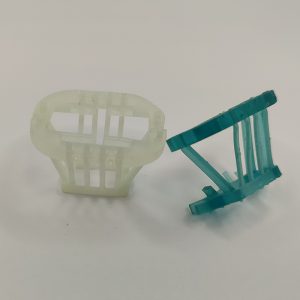
Similarly to the Buzsáki lab head gear, the 3Dneuro crown was developed to simplify implant surgeries. It consists of a flexible base and walls, and is open on the back. This allows it to be placed around an already implanted microdrive. However, if you want to implant more than one drive, you might decide to implant it first to make sure the crown fits around your drives. Two holding braces are then glued on the top of the crown. They provide additional stability. The Omnetics connector is then clamped into one holding brace and fixed with glue or dental acrylic. This results in a slightly less flexible and stable solution for the Omnetics than the mouse caps, but does not require any additional soldering during surgery.
For more information on the 3Dneuro crown, get in touch.
Comparing weight, the copper mesh approach can result in the lightest implant (you can trim and shape the mesh to have the minimal size required for your drive placement), making it most suited for many-drive implants. The rat cap is, unsurprisingly, the heaviest head gear, with the 3D printed parts weighing around 8.3g (with lid, 6.3g without). It is followed by the mouse cap (~1.6g for parts, ~2.2g complete implant) and 3Dneuro crown (~1g for printed parts).
Overview of some head gear systems
| Rat cap base & copper mesh | Rat cap | Mouse cap | 3DNeuro mouse crown | |
|---|---|---|---|---|
| Intended species* | Mouse + Rat | Rat | Mouse* | Mouse* |
|
Difficulty of use
|
Advanced
|
Medium
|
Medium
|
Medium
|
|
Flexibility
|
Extremely flexible
|
Flexible (large enough for most use cases)
|
Limited (space becomes issue with >1 drive)
|
Limited (space becomes issue with >1 drive)
|
|
Time requirement during surgery**
|
20-30min (mouse), 45-60min (rat)**
|
5-10min**
|
5-10min**
|
5-10min**
|
|
Requires soldering during surgery
|
Yes
|
Yes
|
Yes
|
No
|
|
Integrated head-fixation
|
No
|
No
|
No
|
Optional
|
|
Available area***
|
Whole skull
|
 |
 |
 |
|
Approx. Weight (printed parts only) ****
|
0.9g
|
8.3g
|
1.575g
|
1.05g
|
References
[1] Vandecasteele, M., M., S., Royer, S., Belluscio, M., Berényi, A., Diba, K., Fujisawa, S., Grosmark, A., Mao, D., Mizuseki, K., Patel, J., Stark, E., Sullivan, D., Watson, B., & Buzsáki, G. (2012). Large-scale Recording of Neurons by Movable Silicon Probes in Behaving Rodents. Journal of Visualized Experiments, 61. https://doi.org/10.3791/3568
[2] Marzban, N., Anderson, P., & Cohen, M. X. (n.d.). Resonance in prefrontal-striatal circuitry induced by optogenetic activation of ascending dopamine fibers—12th FENS Forum of Neuroscience. Fens 2020 Virtual Forum. Retrieved June 1, 2021, from https://cslide.ctimeetingtech.com/fens2020/attendee/eposter/poster/4334
[3] Mishra, A., Marzban, N., Cohen, M. X., & Englitz, B. (2020). Dynamics of neural microstates in the VTA-striatal-prefrontal loop during novelty exploration in the rat [Preprint]. BioRxiv. https://doi.org/10.1101/2020.08.27.270249
[4] Vöröslakos, M., Petersen, P. C., Vöröslakos, B., & Buzsáki, G. (2021). Metal microdrive and head cap system for silicon probe recovery in freely moving rodent. ELife, 10, e65859. https://doi.org/10.7554/eLife.65859
Last update: June 2, 2021